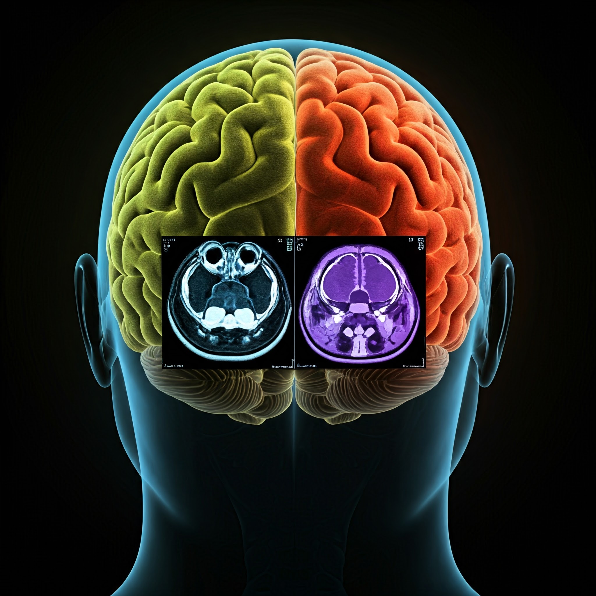
Brain imaging has revolutionized our understanding of the human brain, enabling scientists and researchers to study its structure, function, and underlying mechanisms with unprecedented precision. In recent years, the field of neuroscience has seen remarkable advancements through multimodal brain imaging data and analytics, which offer more comprehensive insights than traditional single-modality approaches. But what exactly is multimodal brain imaging, and why is it so important?
Understanding Multimodal Brain Imaging
Multimodal brain imaging refers to the use of different imaging techniques in a complementary manner to study the brain’s structure and function. By combining multiple types of imaging data, researchers can obtain a richer and more detailed view of brain activity and anatomy than when using a single imaging method. Each imaging modality has its unique strengths, and when integrated, they provide a more holistic picture of brain processes.
Common Imaging Modalities Used
To better understand multimodal brain imaging, let’s explore some of the primary imaging techniques that are often combined:
- MRI (Magnetic Resonance Imaging):
- Used for structural imaging to visualize the anatomy of the brain.
- Offers high spatial resolution and detailed images of brain structures.
- fMRI (Functional MRI):
- Measures changes in blood flow and oxygenation to track brain activity in real-time.
- Ideal for mapping functional brain areas involved in specific tasks.
- PET (Positron Emission Tomography):
- Provides insights into metabolic processes and can trace the activity of specific substances in the brain.
- Used for studying neurochemical changes and mapping brain function.
- EEG (Electroencephalogram):
- Records electrical activity from the brain, providing high temporal resolution.
- Useful for examining the brain’s electrical patterns and real-time activity.
- MEG (Magnetoencephalography):
- Measures the magnetic fields produced by neuronal activity.
- Offers excellent temporal resolution, ideal for understanding brain dynamics over milliseconds.
Why Use Multimodal Brain Imaging?
The combination of these imaging modalities provides a comprehensive approach to understanding brain function and structure. Here’s why multimodal brain imaging data and analytics are important:
- Enhanced Accuracy and Precision:
- By integrating multiple imaging techniques, researchers can cross-verify and complement findings. For instance, fMRI provides excellent spatial information but lacks temporal resolution, while EEG offers high temporal resolution but lower spatial accuracy. Combining both allows for better accuracy in studying brain activity over time and location.
- Comprehensive Analysis:
- Multimodal imaging provides insights that single-modality approaches cannot. For example, combining MRI and PET allows scientists to study both the structural and metabolic aspects of the brain simultaneously.
- Improved Diagnosis and Treatment:
- In clinical settings, multimodal imaging helps diagnose conditions like epilepsy, Alzheimer’s disease, and psychiatric disorders with higher precision. Treatments can be tailored based on a comprehensive understanding of the brain’s condition and how different regions interact.
- Research and Innovation:
- Multimodal imaging drives advancements in cognitive neuroscience, neurology, and psychology by enabling researchers to probe deeper into the complexities of brain function, study the brain’s response to stimuli, and understand neuroplasticity.
Applications of Multimodal Brain Imaging
The use of multimodal brain imaging data and analytics spans several fields, from medical diagnostics to cognitive research. Below are some of the most significant applications:
- Neurodegenerative Disease Studies:
- Multimodal imaging is instrumental in understanding and diagnosing neurodegenerative diseases such as Alzheimer’s and Parkinson’s. By combining structural MRI with PET or SPECT (Single Photon Emission Computed Tomography), researchers can identify structural and functional changes in the brain.
- Brain-Computer Interfaces (BCIs):
- Combining EEG with fMRI can be used to develop BCIs that interpret brain activity and enable interaction with external devices. This can be life-changing for individuals with severe physical disabilities.
- Cognitive and Behavioral Research:
- Using fMRI and EEG together can help scientists study cognitive functions like memory, decision-making, and problem-solving. This is crucial for understanding how different brain areas contribute to these processes and how they are affected in conditions like ADHD or autism.
- Clinical Applications:
- Multimodal imaging allows for personalized treatment plans. For example, understanding how different brain regions interact can help clinicians create targeted therapy strategies for patients with stroke or traumatic brain injuries.
Challenges in Multimodal Brain Imaging
Despite its numerous advantages, multimodal brain imaging comes with certain challenges:
- Data Integration:
- One of the main challenges is aligning data from different modalities with varying resolutions and coordinate systems. This requires sophisticated software tools and algorithms that can fuse data seamlessly for comprehensive analysis.
- Cost and Complexity:
- Multimodal imaging is more expensive and technically complex compared to single-modality studies. The cost of operating various imaging equipment and processing data from these machines can be significant.
- Data Overload:
- The sheer volume of data generated from multimodal imaging studies can be overwhelming. Advanced computational tools and data analysis algorithms are essential to make sense of this information.
Future of Multimodal Brain Imaging
The future of multimodal brain imaging is promising, thanks to advancements in computational power, AI-driven analytics, and innovative imaging technologies. Machine learning models and AI algorithms are being developed to improve data processing, enhance image fusion techniques, and extract meaningful patterns from complex brain data.
Moreover, with the rise of collaborative research networks, the integration of multimodal imaging across various research institutions is paving the way for global studies that can lead to breakthroughs in understanding mental health, brain disorders, and overall cognitive functions.
Conclusion
Multimodal brain imaging is a powerful tool that leverages multiple imaging techniques to gain a more detailed understanding of brain structure and function. By using multimodal brain imaging data and analytics, researchers can gain deeper insights that are essential for advancing neuroscience, improving medical diagnoses, and developing new treatment strategies. While it comes with its challenges, the potential for groundbreaking discoveries makes it an indispensable part of modern neuroscience.
For more information on how multimodal imaging and analytics are shaping the future of neuroscience, stay tuned to Akridata and explore how cutting-edge data analysis solutions are transforming brain research.


No Responses